脳卒中センタ-(SCU6床)/脳神経外科
脳(頭部)に限定されるのではなく
脊椎・脊髄、末梢神経までトータルに対応する「神経外科」です
脳神経外科は英語でニューロサージェリー(neurosurgery)という呼称で、この言葉が意味するのはニューロ=神経、サージェリー=外科で、「神経外科」となります。何故か日本の医療では「脳外」と略称されてしまい、脳神経外科医=頭の医者という誤ったイメージが広まってしまいました。
海外では脳と脊髄および末梢神経も脳神経外科医が外科的治療に携わります。そして頭部の専門医、脊髄末梢神経の専門医はおおよそ同数となります。
当科は、頭部(首から上)だけでなく脊椎、末梢神経にも対応しており、このための医師・スタッフの教育を行っています。
従来より、脊椎脊髄外科治療について日本では、脳外科側の脊髄外科学会と整形外科側の脊椎脊髄外科学会の2つの大きな団体があり、それぞれが認定する専門医制度を行っていたのですが、昨年度より日本専門医機構により脊椎脊髄外科専門医として統一されることになりました。医学は専門分化していますが、患者さんの身体は一つですので、治療する医療者側が疾患や病態に向けて統合されるのは自然の流れと言えるかもしれません。所属する科が重視されるのではなくなってきていると感じています。
当科ではこの本質的な部分を追求し、“患者さんが良くなる”ために何をすべきかを第一に、質を高めていく努力を行っています。
患者さんの負担ができるだけ少なく
できるだけ早い家庭復帰が出来ることを治療の最大の目標にしています
当科では、この20年間にわたり、従来からの開頭外科的手術、脊椎・脊髄と末梢神経の手術、そしてカテーテルを使った血管内治療を3本の柱として、疾患と患者さんの状況にあった適切な治療の提供につとめています。
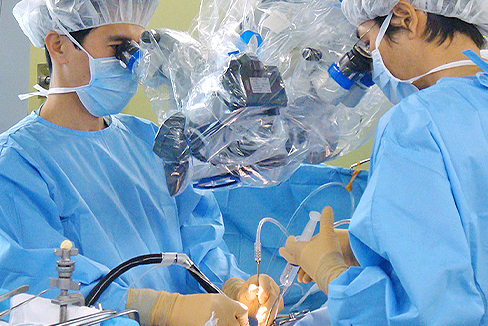
具体的な例で順に説明しますと、開頭や首の切開など、直接目で見て顕微鏡を使って治療するのは最も得意とするところですが、何でもかんでも切るのではありません。
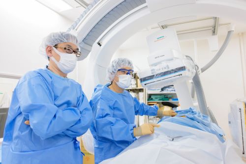
従来の開頭手術のように頭部にメスを入れることなく、主に大腿動脈からカテーテルを脳内や顔面の末梢血管まで挿入して行う血管内手術を積極的に行なっており、非常に困難な症例に関しても良好な結果が得られています。特に、施設・術者限定にしか供給されない一部の頭蓋内ステントや液体塞栓物質などを用いての治療も対応しております。血管内治療で治療可能な疾患は第一選択としています。なお、血管内手術で治療できる主な疾患には以下のものがあります。
- 脳動脈瘤破裂によるくも膜下出血
- 未破裂脳動脈瘤
- 脳・脊髄動静脈奇形
- 脳塞栓症(経皮的血栓回収術的などによる再開通療法)
- 頚部内頚動脈狭窄症
- 脳内主幹動脈の狭窄病変
- 眼球突出や結膜の充血・複視の原因となる内頚動脈海綿静脈洞瘻
- 脳腫瘍や顔面腫瘍の術前栄養血管塞栓
- 外傷(難治性鼻出血、難治性慢性硬膜下血腫)
脊椎の治療も同様です。従来は大きく切開して、患部がよく見える状況にして切除して、固定するというのが一般的な治療でしたが、現在ではできるだけ切開する範囲を狭くし、可能な限り患者さんへのダメージを減らして修復していくことを治療の方針としています。
背骨は体にとって動く柱ですから、削る範囲をできるだけ小さくしてグラグラしないようにします。このことによってネジで固定することを減らすことができるわけです。どうしてもネジを入れる必要がある場合でも、できるだけ筋肉を切らずに治療を行います。背中の筋肉は腰痛に関連しており、これを切ると、回復する期間が長くなるからです。当院では脊椎手術後は、翌日より起立歩行可能で、抜糸後退院となります。高齢者に多い圧迫骨折の場合は骨に医療用のセメントを注入する「経皮的椎体形成術」を行うことで、できるだけ早く痛みをとって早く家庭復帰できるようにしており、当日ないし翌日退院が可能です。
このように、いずれの治療法についても、患者さんの負担ができるだけ少なくし、スムーズな社会・家庭復帰が出来るよう「低侵襲」な医療となることを目指しています。
概要
当科は、さまざまな脳・脊髄神経疾患の症状に応じた検査・治療を実施しています。
診療方針
頭痛、めまい、物忘れなどはもとより 顔手足の痛み、手足のしびれ、筋力低下、歩行障害、腰痛、背部痛、下肢痛などの症状につき治療を行っています。これらの訴えに応じた最も適切な検査を選択し、インフォームドコンセント・セカンドオピニオンを重視し、患者さん・ご家族の同意のもとに、治療方針を決定しています。脳卒中に対しては専門の脳卒中ケアユニットを併設し、救急隊の要請はもとより、近隣の開業の先生方との“病診連携”により、くも膜下出血、脳出血、脳梗塞、頭部脊椎外傷など緊急を要する依頼に対し、SCUホットラインを設けて、24時間、365日、脳外科医が対応可能な体制を採っています。
当施設の特徴
当施設は日本脳神経外科学会専門研修プログラム連携施設(基幹施設番号53 施設番号276号)及び日本脳脊髄外科学会認定訓練施設(21号)です。また、急性期脳梗塞に対するt-PA,血栓粉砕治療実施の認可施設です。圧迫骨折に対する風船治療の認可施設です。
診断機器
診断機器はCT、3DCT、MRI、超音波、脳血管撮影装置、パーフュージョンCT血流動態検査、電気生理学的検査(SEP、ABR、NCV)などを完備しています。脳神経専門の検査技師、放射線科技師と密接に症例検討を行っております。
脳血管撮影装置は最高機種のバイプレーンフラットパネルを導入しており、超微細な脳血管内治療が可能となっています。また、手術室には回転Cアームとナビゲーションシステムをもち、手術を低侵襲、安全に実施可能となっています。脊椎疾患や末梢神経疾患は当院神経筋外来の協力のもと電気生理学的検査(SEP、NCV)などによる客観的評価を必須とし、また脳血管障害はすべて専門技師により超音波を用いた全身の脈管学的精査フォローを行っています。
主な治療対象疾患は、脳腫瘍、脊髄腫瘍、くも膜下出血、未破裂脳動脈瘤、脳動静脈奇形、脳内出血、脳梗塞等の脳血管障害、急性硬膜外・下血腫、脳挫傷、慢性硬膜下血腫等の頭部外傷、脊髄損傷、頸椎症、頸椎椎間板ヘルニア、腰部脊柱管狭窄症、腰椎すべり症、圧迫骨折、腰椎椎間板ヘルニアなどの脊椎疾患、手根管症候群、肘部管症候群などの末梢神経疾患、顔面けいれん、三叉神経痛などの機能的疾患など脳神経外科全般にわたります。
また、従来の開頭手術のように頭部にメスを入れることなく、大腿動脈からカテーテルを脳内の血管や顔面の血管まで挿入して行う新しい治療手段“血管内手術”により、非常に困難な症例に関しても良好な結果が得られています。血管内治療で治療可能な疾患は第一選択としています。尚、血管内手術で治療できる主な疾患には以下のものがあります。
- 脳動脈瘤破裂によるくも膜下出血
- 未破裂脳動脈瘤
- 脳・脊髄動静脈奇形
- 脳塞栓症(急性期血栓溶解療法)
- 脳梗塞の原因となる動脈硬化性狭窄病変
- 眼球突出や結膜の充血
- 複視の原因となる頚動脈海綿静脈洞瘻
- 脳腫瘍や顔面腫瘍の術前栄養血管塞栓
脊椎圧迫骨折に対しては、経皮的椎体形成術(セメント治療)を平成15年7月から実施し600症例を超えました。除痛効果に優れ、日帰り治療が可能です。
診療体制
診療内容の評価
外科診療にとりもっとも大きなウェートを占めるのは手術成績であり、成績・手技向上のため、手術全過程をビデオに記録保存、デジタル化することにより、論文、学会、他施設との症例検討会などを通じて公表、他者評価を受けるようにしています。また、後輩やパラメディカルの指導に用いています。手術症例は全てデータベース化し、検索、集計、発表ができるようにしています。
教育・研究・治験
論文発表・学会活動を通じて他者批判を乞うとともに、最先端の治療行為は他大学や外国での遺体解剖による手術訓練を行い、研究所での動物実験にて修練を積んでいます。また後進の教育方針としては、開頭手術、脊椎・末梢神経手術、血管内手術を3本の柱としていずれにも対応できる脳神経外科専門医を養成しております。手術室以外での顕微鏡手術修練(off job)を行っています。具体的には以下のプログラムを実施しています。
【臨床研修年次別プログラム(診断、検査、患者管理、手術など)】
- 1年次 救急外来処置、ICU管理、腰椎穿刺、脊髄造影、脳血管撮影の助手術者、穿頭術、顕微鏡手術操作の修練
- 2年次 開頭術・脊椎手術の術者助手、脳室ドレナージ
- 3年次 急性硬膜下血腫、急性硬膜外血腫、VPシャント術
- 4年次 開頭脳内血腫除去術、頸椎椎弓形成術、などの術者選択的血栓溶解術の術者
- 5年次 STAMCA吻合術、腰椎椎間板ヘルニア摘出術、経皮的血管形成術、外頸動脈塞栓術
- 6年次以降 脳動脈瘤クリッピング術、脳腫瘍摘出術、頸椎前方固定術、動脈瘤塞栓術
【カンファレンスなど】
- 月曜日AM 8時から 英文抄読会
- 火曜日AM 8時から 臨床カンファレンス
- 金曜日AM 8時から 臨床カンファレンス
当診療科では、下記事業に参加しています。詳しくは、以下をクリックして下さい。
脳卒中とSCUについて
脳卒中は、脳の血管が詰まる脳梗塞、高血圧などにより血管が切れる脳出血、特に脳動脈瘤が破裂するクモ膜下出血、に分類されます。降圧・脂質管理を主とした予防医療の進歩により脳出血は減少しました。しかしクモ膜下出血は減少しておらず、脳梗塞はむしろ増加している現状です。
脳卒中は、がん・心臓病に次いで死因第3位であり、年間13万人くらいの方が亡くなっています。寝たきりになる原因の3割近くを占め、第1位です。高齢化社会や生活習慣病の増加もあり、毎年25万人以上が新たに発症されています。全国の患者数は2020年には250万人、約60%が要介護と試算されています。
このような社会背景に適した脳卒中の対応が望まれます。つまり、脳卒中に対して予防治療から急性期・亜急性期・慢性期治療、在宅医療まで統合的・系統的な対応が必要と考えられます。これには、医師、看護師、リハビリテーション療法士などの医療従事者および医療ソーシャルワーカーによるチーム医療、近隣のリハビリ病院、掛かり付け医、介護施設などとの連携の整備が不可欠です。
脳卒中ケアユニット(stroke care unit:SCU)とは、脳卒中急性期の患者さんを専門医療スタッフが、急性期の集中治療とリハビリテーションとを組織的、計画的に行う脳卒中専用の治療病棟のことです。脳卒中治療ガイドライン(2009)では、SCUで治療することによって、脳卒中患者の死亡率の低下、在院期間の短縮、自宅退院率の増加、長期的な日常生活動作(ADL)と生活の質(QOL)の改善を図ることができるという検証結果が示されました(グレードA)。
当科ではこのような社会・地域の要望にこたえるために、厚生労働大臣の定める施設基準を満たすSCUを6床開設、脳卒中センターを設立し、脳卒中急性期の受入れ・治療を今まで以上に充実させて頂きました。脳卒中専門医を含む脳神経外科または神経内科の経験あるスタッフが24時間対応致します。脳卒中の可能性の高い症状を示す患者さんはできる限り早くご連絡下さい。脳卒中かどうかの判断が困難な患者さんも遠慮、躊躇なくご相談下さい。
尚、SCUおよび脳神経外科の病床数は限られております。治療・診療経過に応じて、更なるリハビリが必要な患者さんには地域連携パスによる回復期リハビリ病院への転院、その他の介護施設・療養型病院などへの転院マネージメントも行わせて頂いております。
研究・治験
将来計画
患者さんの負担の少ない医療の推進:当科で積極的に進めている脳血管内治療はもちろんのこと、従来の外科治療を常に再検討し、最先端の医療を柔軟に選択していくことが必要と考えています。脊椎圧迫骨折に対する経皮的椎体形成術(セメント治療)を平成15年7月から導入し600症例を超えました。
セメント治療は当施設が学会事務局となり、安全な治療となるべく、学会を主宰しています(*)。なお圧迫骨折に対するセメント治療は、最新医学新書(最新医学社)「腰痛治療の最前線―圧迫骨折はセメントで直せー」にて紹介しております。
また脊椎手術は従来のように大きくきるのではなく、顕微鏡やナビゲーションを駆使して、小さな傷で行い、痛みを極力減らす努力をしています。手術翌日には全例歩行が可能です。
(*)椎体形成術研究会
事務局 医仁会 武田総合病院 脳神経外科 川西昌浩
クリニカルパス
About us
Neurosurgery Services & Treatment
In Japan, most neurosurgery facilities only perform brain surgery, while spine and peripheral nerves are handled by orthopedic surgeons. The Department of Neurosurgery at Takeda General Hospital provides total treatment not only for the brain, but also for the spinal cord and peripheral nerves. All staff members in our department are striving to improve the quality of our services with a focus on what we need to do to "help patients get better.
Our Goal
Our main goal is to help patients return home as soon as possible with as little burden as possible.
For the past 25 years, we have been providing appropriate treatment for each disease and each patient's situation by equating traditional craniotomy, spine and peripheral nerve surgery, and endovascular treatment with catheterization.
We are actively performing endovascular surgeries in which catheters are inserted into peripheral vessels in the brain and face, mainly through the femoral artery, without cutting the head as in conventional craniotomy, and have obtained excellent results even in very difficult cases. Using the latest angiography equipment, only one of which has been introduced in Kyoto, we are also able to provide treatment using some intracranial stents and liquid embolization materials, which are supplied only to facilities and surgeons. Diseases that can be treated by endovascular therapy are our first choice.
Our Philosophy
In the past, the treatment of the spine was to make a large incision, remove the affected area with a clear view, and fix it. However, our current treatment policy is to make the incision as small as possible and to use a microscope to repair the area with as little damage to the patient as possible.
Since the spine is a moving pillar of the body, our concept is to keep the area of removal as small as possible to avoid wobble. This reduces the need for screws to hold it in place. Even when it is necessary to insert screws, the treatment is performed with as little cutting of the muscles as possible. This is because back muscles are associated with back pain, and cutting them will prolong the recovery period. After spinal surgery at our clinic, patients are able to stand and walk from the next day and are discharged after stitches are removed. For compression fractures, which are common among the elderly, we perform "percutaneous vertebroplasty," in which medical cement is injected into the bone, to relieve pain as quickly as possible and allow patients to return home as soon as possible, allowing them to leave the hospital the same day or the day after.
In this way, we aim to provide "minimally invasive" medical care that minimizes the burden on the patient and allows for a smooth return to society and home.
Service
The main diseases that can be treated by endovascular surgery include the following.
- Unruptured cerebral aneurysm
- Subarachnoid hemorrhage due to ruptured cerebral aneurysm
- Cerebral and spinal cord arteriovenous malformations
- Cerebral embolism (percutaneous thrombus retrieval and other recanalization procedures)
- Stenosis of the internal carotid artery in the neck
- Stenotic lesions of the main intracerebral arteries
- Internal carotid cavernous sinus fistula causing eye protrusion, chemosis, and diplopia
- Preoperative feeding vascular embolization of brain and facial tumors
- Trauma (intractable epistaxis, intractable chronic subdural hematoma)
Major diseases
Major diseases treated include brain tumors, spinal cord tumors, cerebrovascular disorders such as subarachnoid hemorrhage, unruptured cerebral aneurysm, cerebral arteriovenous malformation, intracerebral hemorrhage, cerebral infarction, head injury such as acute epidural and subdural hematoma, cerebral contusion, chronic subdural hematoma, spinal cord injury, cervical spondylolisthesis, cervical disc herniation, lumbar spinal canal stenosis, lumbar spondylolisthesis, compression fracture, lumbar disc herniation, and other spinal disorders, peripheral nerve diseases such as carpal tunnel syndrome, elbow tunnel syndrome, and functional disorders such as facial spasm and trigeminal neuralgia. Spinal disorders such as hernias, peripheral nerve disorders such as carpal tunnel syndrome and elbow tunnel syndrome, functional disorders such as facial spasm and trigeminal neuralgia, and other general neurosurgical conditions.
Percutaneous vertebroplasty
Percutaneous vertebroplasty (cement treatment) has been performed for spinal compression fractures since July 2003, and more than 1000 cases have been treated.It has excellent pain-relieving effects and can be performed as a one-day treatment.
https://www.e-neurospine.org/journal/view.php?number=1488
DOI: https://doi.org/10.14245/ns.2346936.468
Our angiography and fluoroscopy system (ARTIS icono D-Spin, Siemens)
What are the features of this machine?
- 1. No complicated operation is required for 3D imaging,
- 2. It has an excellent memory function for device position information, just like a navigation system,
- 3. Reduced fluoroscopic (i.e., radiation exposure) dose
- 4. Shortening of examination and operation time
- 5. Rapid analysis and simulation of virtual stents, etc.
- 6. Multifaceted consideration is possible in combination with metabolic (cerebral blood flow) evaluation.
In other words, in addition to "visualization," simple and rapid "analysis" is possible, which reduces the burden on the physician performing the procedure and, by extension, has a positive effect on patient care.
In addition, the so-called "hybrid operating room" has been completed and put into operation with cleanliness control equivalent to that of an operating room. In the past 10 years, we have witnessed the advent of the "intracranial stent era," and the structures are becoming more delicate.
Stent Therapy Treatment of Cerebral Aneurysms
The treatment of cerebral aneurysms with stents is becoming increasingly popular, and there are two main types of stents.
The general stent has a rough mesh structure and its main purpose is to act as a bulwark to prevent the coils to be embolized in the aneurysm from protruding into the mother vessel.
On the other hand, a flow diverting stent is a tool that reduces blood flow into the aneurysm and causes thrombosis by placing a stent with finer stents into the mother vessel.
Of course, not all aneurysms can be treated with all of these tools, as they are not suited to all patients.
When treating aneurysms with the FLOW DIVERTER, we are able to perform the procedure with the help of "Verify Now," a testing device that can check platelet aggregation (blood thinning), and with an imaging device with operability and analysis capabilities, such as our department's angiography and fluoroscopy equipment (ARTIS icono D-Spin, Siemens). We are confident in our ability to provide "world standard" treatment to patients in the region.What are the features of this machine?
- 1. No complicated operation is required for 3D imaging,
- 2. It has an excellent memory function for device position information, just like a navigation system,
- 3. Reduced fluoroscopic (i.e., radiation exposure) dose
- 4. Shortening of examination and operation time
- 5. Rapid analysis and simulation of virtual stents, etc.
- 6. Multifaceted consideration is possible in combination with metabolic (cerebral blood flow) evaluation.
In other words, in addition to "visualization," simple and rapid "analysis" is possible, which reduces the burden on the physician performing the procedure and, by extension, has a positive effect on patient care.
In addition, the so-called "hybrid operating room" has been completed and put into operation with cleanliness control equivalent to that of an operating room. In the past 10 years, we have witnessed the advent of the "intracranial stent era," and the structures are becoming more delicate.
Physicians and areas of focus
| |
| |
| |
| |
| |
| |
|
Evaluation of Medical Practice
In order to improve surgical outcomes and techniques, the entire surgical process is recorded on video and digitized so that it can be published and evaluated by others through papers, academic conferences, and case review meetings with other institutions. We also use this information to provide guidance to junior surgeons and para-medical staff. All surgical cases are stored in a database so that they can be searched, tabulated, and published.
Office Hours
working day | Mon-Fri. 9;00-12;00 |
Address | 28-1 Ishidamori minamimachi Fushimiku Kyoto city Kyoto |
Toll Free | 0120-72-6530 |
Tel. | +81-75-572-6530(Direct line) |
Fax. | +81-75-572-6276(Direct line) |
Secretary | Tsujino SW |
Access | Kyoto Metro Tozai line Ishida Sta. Exit 1. 5 minutes walk |
Map |
Evaluation of Medical Practice
In order to improve surgical outcomes and techniques, the entire surgical process is recorded on video and digitized so that it can be published and evaluated by others through papers, academic conferences, and case review meetings with other institutions. We also use this information to provide guidance to junior surgeons and para-medical staff. All surgical cases are stored in a database so that they can be searched, tabulated, and published.
Education, Research, and Clinical Trials
We receive criticism from others through publications and academic conference activities, and we conduct surgical training through autopsies of cadavers at other universities and in foreign countries for cutting-edge therapeutic acts, and we conduct animal experiments at our research institute. In addition, we train neurosurgeons who can handle craniotomy, spine/peripheral nerve surgery, and endovascular surgery as the three pillars of our education policy for future generations. We offer off-job training in microsurgery outside of the operating room.
Conferences, etc.
Our department participates in the following activities. For more information, please click below.
About Stroke and SCU
Stroke is classified into three types: cerebral infarction, in which a blood vessel in the brain becomes blocked; cerebral hemorrhage, in which a blood vessel breaks due to high blood pressure or other causes; and subarachnoid hemorrhage, in which a cerebral aneurysm ruptures. Advances in preventive medicine, mainly in lowering blood pressure and lipid management, have reduced the incidence of cerebral hemorrhage. However, subarachnoid hemorrhage has not decreased, and cerebral infarction is on the increase.
Stroke is the third leading cause of death after cancer and heart disease, killing about 130,000 people annually. It is the leading cause of death, accounting for nearly 30% of those who become bedridden. More than 250,000 new cases are diagnosed each year, due in part to the aging of the population and the increase in lifestyle-related diseases. It is estimated that the number of patients nationwide will reach 2.5 million by 2020, with approximately 60% requiring long-term care.
It is desirable to respond to stroke in a way that is appropriate for such a social background. In other words, an integrated and systematic response to stroke, from preventive treatment to acute, subacute, and chronic treatment, as well as home care, is considered necessary. To this end, it is essential to establish a team of medical professionals including physicians, nurses, rehabilitation therapists, and medical social workers, as well as cooperation with nearby rehabilitation hospitals, primary care physicians, and nursing care facilities.
It is desirable to respond to stroke in a way that is appropriate to this social context. In other words, an integrated and systematic response to stroke is considered necessary, from preventive treatment to acute, subacute, and chronic treatment, as well as home care. To this end, it is essential to establish a team of medical professionals including physicians, nurses, rehabilitation therapists, and medical social workers, as well as cooperation with neighboring rehabilitation hospitals, primary care physicians, and nursing care facilities.
A stroke care unit (SCU) is a specialized treatment ward for acute stroke patients where specialized medical staff provides intensive acute treatment and rehabilitation in an organized and systematic manner. The Stroke Treatment Guidelines (2009) verified that treatment in the SCU can reduce mortality, shorten hospital stay, increase the rate of discharge home, and improve long-term activities of daily living (ADL) and quality of life (QOL) of stroke patients (Grade A).
In order to respond to such social and community demands, the Department opened a 6-bed SCU that meets the facility standards specified by the Minister of Health, Labor and Welfare, and established the Stroke Center to better accept and treat acute stroke patients than ever before. Experienced neurosurgeons and neurologists, including stroke specialists, are available 24 hours a day. Patients with symptoms that indicate a high probability of stroke should contact us as soon as possible. Patients who have difficulty in determining whether or not they are suffering from a stroke should not hesitate to contact us.
The number of beds in the SCU and Neurosurgery Department is limited. Depending on the course of treatment and medical care, patients who require further rehabilitation may be transferred to a convalescent hospital through a regional cooperation path, or managed for transfer to other long-term care facilities or convalescent hospitals.
Research and Clinical Trials
Our medical treatment is widely publicized through papers, video presentations, and academic conferences, both in English and Japanese.
外来診療表
※都合により外来担当医等が変更している場合がございますので、以下の◎休診・代診情報をご覧頂くか、或いは直接お電話にてお問合わせの上ご確認頂きますようお願い致します。
脳卒中センター(脳神経外科)
月 | 火 | 水 | 木 | 金 | 土 | |
---|---|---|---|---|---|---|
午前 | 予約優先田中 秀一
|
予約優先横山 邦生
|
予約優先川西 昌浩
|
予約優先山田 誠
|
予約優先伊藤 裕
|
|
午後 | 予約のみ山田 誠
(脳血管障害・血管内治療専門外来
13時~15時) |
予約優先池田 直廉
|
令和5年10月より、土曜日午前診につきましては閉診となります。
大変ご迷惑をお掛け致しますが、ご理解とご協力をお願い致します。
休診・代診情報
- 2024年10月18日(金) 脳卒中センター(脳神経外科) (午後):池田→休診
- 2024年11月08日(金) 脳卒中センター(脳神経外科) (午後):池田→休診
予約外来
当院では待ち時間短縮のため、下記の印の科目については予約制を導入しています。
- 予約優先・・・初診・予約のない再診・急患の方も受付けています。
- 予約のみ・・・予約をされた再診の方のみの診療となっています。(初診の方は受け付けておりません。)
- 電話予約可・・・電話で予約を受け付けています。
- 患サポ経由・・・患者サポートセンターからの紹介のみです。